
Presentation
Inflammatory back pain and uveitis. HLA B27 positive.
Patient Data
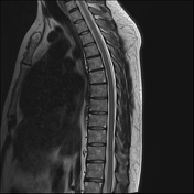

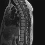
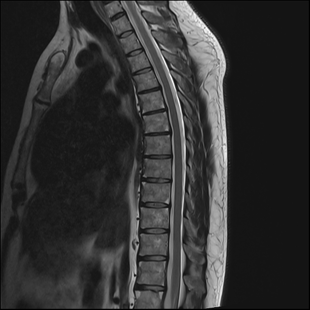
Normal vertebral body alignment. No vertebral body or posterior element ankylosis evident. No syndesmophytes evident.
Several thoracic vertebral bodies demonstrate high T1 and T2 signal corners adjacent to endplates representing Romanus lesions (T5 and T6 are good examples).
Disc spaces are preserved. No disc pathology. No spinal canal or neural foraminal stenosis. The spinal cord, conus, cauda equina and exiting nerve roots are normal.
Patient went on to lumbar spine imaging.


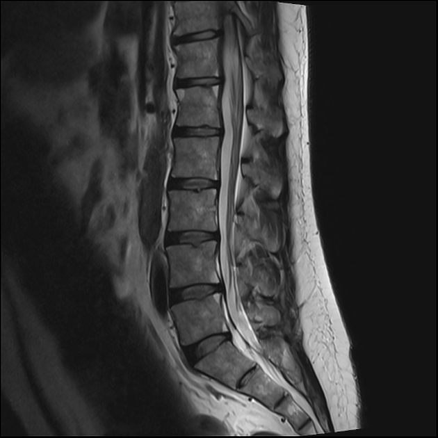
Normal vertebral body alignment. No vertebral body or posterior element ankylosis evident. No syndesmophytes evident.
Several lumbar vertebral bodies demonstrate high T1 and T2 signal corners adjacent to endplates representing Romanus lesions (L1 and L5 are good examples).
Disc spaces are preserved. No disc pathology. No spinal canal or neural foraminal stenosis. The lower spinal cord, conus, cauda equina and exiting nerve roots are normal.
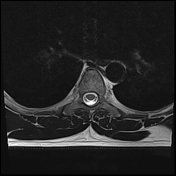
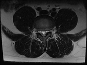
Upper SI joints appear normal.
Impression
The presence of multiple thoracolumbar Romanus lesions and minor sacroiliitis suggest a diagnosis of ankylosing spondylitis. No ankylosis identified in the thoracolumbar spine or upper SI joints.
Case Discussion
The patient was referred to the rheumatology clinic and has responded well to the treatment for ankylosing spondylitis.


 Unable to process the form. Check for errors and try again.