
Locally invasive prostatic cancer with metastatic lymphadenopathies
Presentation
Pelvic pain and hematuria.
Patient Data
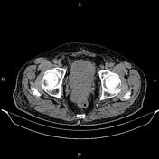

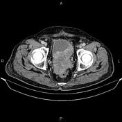

A 100×70 mm mass is present at the anatomical location of the prostate gland that infiltrates the urinary bladder base and the anterior wall of the rectum. The mass also involves the distal portion of the left ureter.
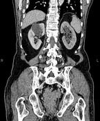
Several lymphadenopathies are noted at bilateral parailiac regions with SAD less than 29 mm. Additionally, several lymphadenopathies are seen at lower para aortocaval regions with a maximum SAD of 18 mm.
A 24 mm thin walled non-enhanced cyst is noted at segment IVa of the liver.
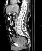
A 60 mm simple cortical cyst with parapelvic extension is noted at the upper pole of the right kidney. Mild to moderate hydroureteronephrosis is seen on the left side.
In imaged portions of the lower thorax, a little volume of pleural effusion is seen at both sides.
Fat containing umbilical hernia is seen.
Case Discussion
Locally invasive pathology proven prostatic adenocarcinoma with abdominopelvic lymphadenopathies.


 Unable to process the form. Check for errors and try again.