
Presentation
Incidental finding in a patient with left flank pain.
Patient Data
Age: 50 years
Gender: Male
From the case:
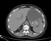
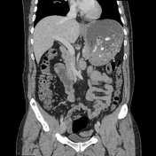
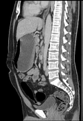
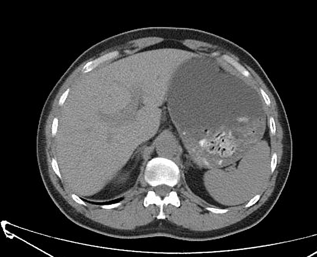
Left-sided IVC with hemiazygos continuation


Download
Info
Left renal stone (7 mm, mean density = 760 HU) in the middle calyx.
Small distal left ureteral stone at the vesicoureteric junction (4 mm, mean density 320 HU) obstructing the ureteric lumen with mild ureteral dilatation.
Interruption of the infrahepatic segment of the IVC with hemiazygos continuation. The retro aortic right renal vein is drained into the dilated hemiazygos vein. The left renal vein is directly drained into a left-sided IVC. The hepatic veins join and drain directly into the supradiaphragmatic IVC.
Case Discussion
CT features of a left-sided IVC with hemiazygos continuation (incidental finding) in a patient with urolithiasis.


 Unable to process the form. Check for errors and try again.