
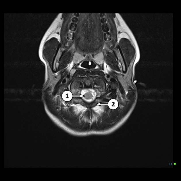
The labeled structures are (excluding the correct side):
cervical spinal cord
posterior arch of C1
odontoid process (peg or dens) of C2
parotid gland
intradural segment (V4) of dominant vertebral artery
cisterna magna
intradural segment (V4) of non-dominant vertebral artery
cerebellar tonsil
occipital condyle
medulla oblongata
lateral foramen (of Luschka)
cerebellar folia
fluid within mastoid air cells
median foramen (of Magendie)
basilar artery
inferior cerebellar vermis
premedullary cistern
pyramid of medulla
inferior cerebellar peduncle
dentate nucleus of cerebellum
internal acoustic (or auditory) canal
cerebellopontine angle cisterna
facial colliculus
nodule of cerebellum
middle cerebellar peduncle
fourth ventricle
Meckel's cave
superior cerebellar peduncle
cavernous segment of internal carotid artery
pons
intraorbital segment of optic nerve
temporal lobe
pituitary gland
falx cerebri
pituitary infundibulum
temporal horn of lateral ventricle
parahippocampal gyrus
inferior colliculus
superior cerebllar vermis
suprasellar cistern
optic chiasm
interpeduncular cistern
cerebral crus (peduncle)
cerebral aqueduct (of Sylvius)
superior colliculus
gyrus rectus
cisternal segment of optic nerve
First segment of middle cerebral artery (M1)
optic tract
mammillary body
substantia nigra
red nucleus
ambient cistern
second segment of posterior cerebral artery (P2b)
basal veins (of Rosenthal)
quadrigeminal plate cistern
pineal gland
occipital horn of lateral ventricle
great cerebral vein (of Galen)
second segment of anterior cerebral artery (A2)
third ventricle
interthalamic adhesion (massa intermedia)
internal cerebral veins
occipital lobe
anterior commissure
Sylvian (lateral) fissure
extreme capsule
insular cortex
claustrum
external capsule
straight sinus
anterior limb of internal capsule
column of fornix
interventricular foramen (of Monro)
posterior limb of internal capsule
thalamus
atrium (trigone) of lateral ventricle
head of caudate nucleus
genu of internal capsule
putamen
globus pallidus
crus of fornix
splenium of corpus callosum
genu of corpus callosum
frontal horn of lateral ventricle
septum pellucidum
forceps major
forceps minor
body of lateral ventricle
choroid plexus
frontal lobe
superior sagittal sinus
body of corpus callosum
centrum semiovale
parietal lobe
postcentral gyrus
central sulcus (of Rolando)
precentral gyrus
interhemispheric fissure
superficial cortical vein

The same normal brain T2 MRI without labels for reference.
Case Discussion
The labeled structures are (excluding the correct side):
cervical spinal cord
posterior arch of C1
odontoid process (peg or dens) of C2
parotid gland
intradural segment (V4) of dominant vertebral artery
cisterna magna
intradural segment (V4) of non-dominant vertebral artery
cerebellar tonsil
occipital condyle
medulla oblongata
lateral foramen (of Luschka)
cerebellar folia
fluid within mastoid air cells
median foramen (of Magendie)
basilar artery
inferior cerebellar vermis
premedullary cistern
pyramid of medulla
inferior cerebellar peduncle
dentate nucleus of cerebellum
internal acoustic (or auditory) canal
cerebellopontine angle cisterna
facial colliculus
nodule of cerebellum
middle cerebellar peduncle
fourth ventricle
Meckel's cave
superior cerebellar peduncle
cavernous segment of internal carotid artery
pons
intraorbital segment of optic nerve
temporal lobe
pituitary gland
falx cerebri
pituitary infundibulum
temporal horn of lateral ventricle
parahippocampal gyrus
inferior colliculus
superior cerebllar vermis
suprasellar cistern
optic chiasm
interpeduncular cistern
cerebral crus (peduncle)
cerebral aqueduct (of Sylvius)
superior colliculus
gyrus rectus
cisternal segment of optic nerve
First segment of middle cerebral artery (M1)
optic tract
mammillary body
substantia nigra
red nucleus
ambient cistern
second segment of posterior cerebral artery (P2b)
basal veins (of Rosenthal)
quadrigeminal plate cistern
pineal gland
occipital horn of lateral ventricle
great cerebral vein (of Galen)
second segment of anterior cerebral artery (A2)
third ventricle
interthalamic adhesion (massa intermedia)
internal cerebral veins
occipital lobe
anterior commissure
Sylvian (lateral) fissure
extreme capsule
insular cortex
claustrum
external capsule
straight sinus
anterior limb of internal capsule
column of fornix
interventricular foramen (of Monro)
posterior limb of internal capsule
thalamus
atrium (trigone) of lateral ventricle
head of caudate nucleus
genu of internal capsule
putamen
globus pallidus
crus of fornix
splenium of corpus callosum
genu of corpus callosum
frontal horn of lateral ventricle
septum pellucidum
forceps major
forceps minor
body of lateral ventricle
choroid plexus
frontal lobe
superior sagittal sinus
body of corpus callosum
centrum semiovale
parietal lobe
postcentral gyrus
central sulcus (of Rolando)
precentral gyrus
interhemispheric fissure
superficial cortical vein


 Unable to process the form. Check for errors and try again.