
Presentation
Young female patient presented with right flank pain.
Patient Data


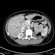
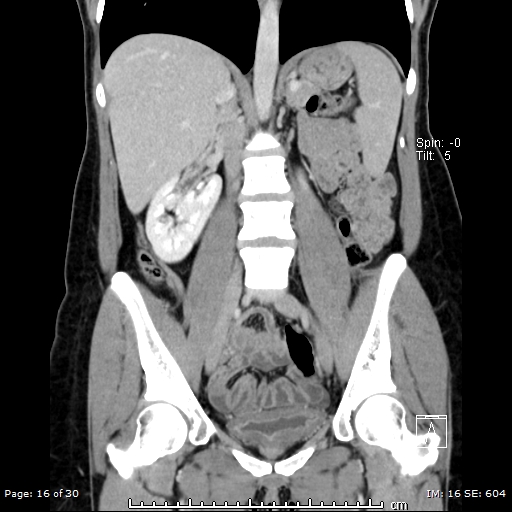
Ectopic left kidney which is seen on the right side, fused with the inferior pole of the right kidney, giving L-shape of the kidney.
Both kidneys appear malrotated, their pelves are pointing anteriorly.
Both kidneys have an extrarenal pelvis.
Bilateral renal arteries arise from the abdominal aorta and bilateral renal veins drain into the IVC.
Features are consistent with crossed fused renal ectopia (type e).
Incidental finding of incomplete annular pancreas (8-3 O'clock), without significant duodenal stenosis.
Case Discussion
In a crossed fused renal ectopic kidney, complications such as nephrolithiasis, infection, and hydronephrosis approaches ~50%.
Crossed fused ectopia usually does not require any primary treatment. The blood supply to the crossed fused kidney is usually anomalous, and angiography is recommended before surgical intervention1.


 Unable to process the form. Check for errors and try again.