Presentation
Left flank pain and fullness on physical exam.
Patient Data
Age: 65 years
Gender: Female
From the case:
Left ureteropelvic junction obstruction
![](https://prod-images-static.radiopaedia.org/images/56387815/110001_thumb.jpeg)
![](https://prod-images-static.radiopaedia.org/images/56387816/110002_thumb.jpeg)
![](https://prod-images-static.radiopaedia.org/images/56387817/110003_thumb.jpeg)
![](https://prod-images-static.radiopaedia.org/images/56387818/220001_thumb.jpeg)
![](https://prod-images-static.radiopaedia.org/images/56387819/220002_thumb.jpeg)
![](https://prod-images-static.radiopaedia.org/images/56387820/33_thumb.jpeg)
![](https://prod-images-static.radiopaedia.org/images/56387821/440001_thumb.jpeg)
![](https://prod-images-static.radiopaedia.org/images/56387822/440002_thumb.jpeg)
![](https://prod-images-static.radiopaedia.org/images/56387834/440003_thumb.jpeg)
![](https://prod-images-static.radiopaedia.org/images/56387848/440004_thumb.jpeg)
Download
Info
![](https://prod-images-static.radiopaedia.org/images/56387815/110001_big_gallery.jpeg)
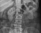
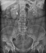
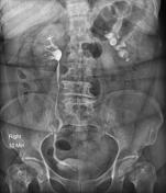
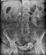
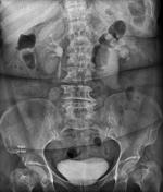
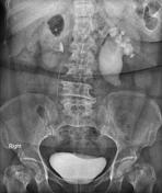
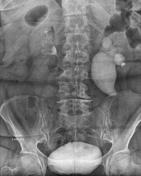
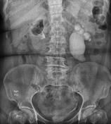
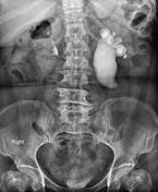
There is left hydronephrosis and delayed contrast excretion with marked pelvic dilatation and normal caliber ureter, suggesting stenosis of the ureteropelvic junction.
Case Discussion
This case demonstrates typical appearances of pronounced left pelviureteric junction obstruction.