
Presentation
A worker presented with an open-cut wound, after injuring his index finger with an angle grinding machine.
Patient Data
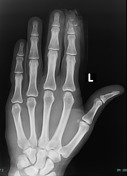
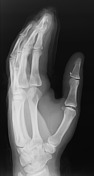

A comminuted displaced open fracture of the tuft and distal shaft of the distal phalanx of the index finger is noted.
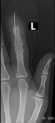
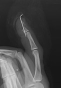
Following debridement of the wound, a K-wire was inserted across the DIP joint into the middle phalanx, to achieve more stable fixation.
A small radiopaque foreign body is seen in the distal thumb.
Case Discussion
Distal phalanx fracture is the most common fracture of the hand, followed by metacarpal bones fracture 1. These fractures can be open or closed.
The diagnosis is based on the history of trauma, mechanism of injury, and x-ray films.
Open fracture of the distal phalanx often necessitates referral to an orthopedic and requires extensive wound debridement, irrigation, soft tissue repair, and antibiotic coverage due to the high risk of infection.
Tetanus booster should be updated, if necessary and surgery for nailbed repair and/or K-wire fixation will be required in complex cases 2.


 Unable to process the form. Check for errors and try again.