
Presentation
Lower limb weakness and loss of sensation for 3 weeks.
Patient Data


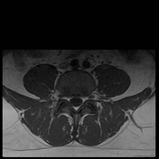
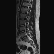
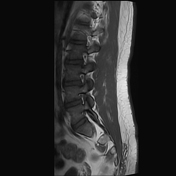

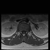
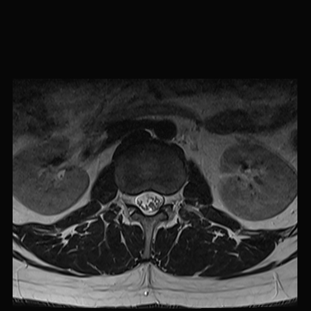
MRI shows a distal conus lesion at L3 demonstrating iso to mid hypointense signal on T1W, T2W hypointense signal and homogenous post-contrast enhancement.
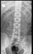
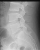
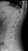

Normal X-ray with degenerative changes or destructive bone lesions
Histopathology:
Macroscopy: specimen labeled "spine tumor in formalin" consists of a well circumscribed mass measuring 20 x 18 x 10 mm.
Microscopy: The sections show a well circumscribed lesion demonstrating variable hypo- and hypercellular areas, composed of a proliferation of bland spindled cells with wavy nuclei. Verocay bodies are present. The background demonstrates thick hyalinised blood vessels and vascular congestion. There is no evidence of necrosis, atypia or malignancy.
Immunohistochemistry:
- S100: diffusely positive in the spindled cells
- Ki67: 2-3%
Pathological diagnosis: spinal cord nerve root, excision: schwannoma.
Case Discussion
The imaging demonstrates an intra-medullary enhancing mass lesion and the patient subsequently underwent surgical resection with histology showing a well-circumscribed hypercellular lesion composed of a proliferation of bland spindled cells with waxy nuclei. Verocay bodies were present and immunohistochemistry was diffusely positive for s100. The pathological diagnosis was consistent with schwannoma.


 Unable to process the form. Check for errors and try again.