
Presentation
CT neck for evaluation of cystic laryngeal lesion on ENT examination. Known case of primary hypothyroidism.
Patient Data


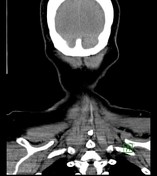
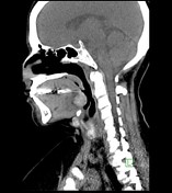
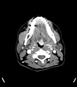
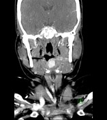
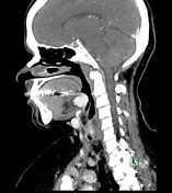
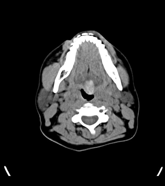
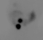
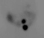
A hyperdense lesion measuring 14 x 22 mm is seen at the midline base of tongue; another similar lesion measuring 11 x 19 mm is seen in the midline neck just below the hyoid bone. Both lesions show intense homogeneous enhancement on post-contrast images. No thyroid tissue is appreciable at its expected normal position.
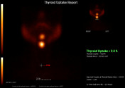
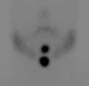
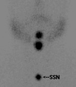
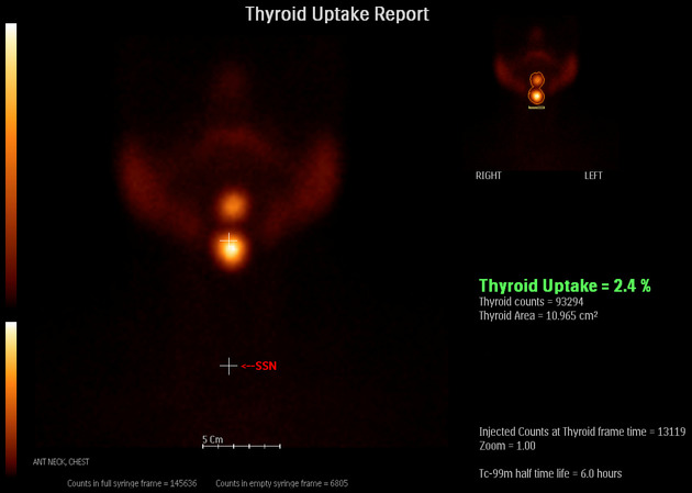
Two focal areas of increased radiotracer concentration lying close to each other, are noted in the midline upper neck, with the superior focus being located along the posterior aspect of the tongue. No functioning thyroid tissue at the expected location of the thyroid gland. Thyroid uptake function: 2.4% (Normal: 0.4-4.0%).
Case Discussion
Non-visualization of the thyroid gland at its expected normal position in the lower neck with finding of two lesions in the neck along the expected migration route of the thyroid gland. These lesions show intense homogeneous enhancement on the CT scan and increased radiotracer uptake on the Tc-99m pertechnetate thyroid scan and are consistent with ectopic thyroid.


 Unable to process the form. Check for errors and try again.