
Presentation
Irregular menstrual period and Increasing abdominal girth .
Patient Data


All standard sequences were obtained.
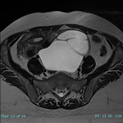
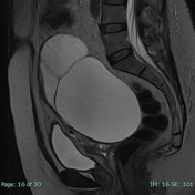
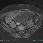
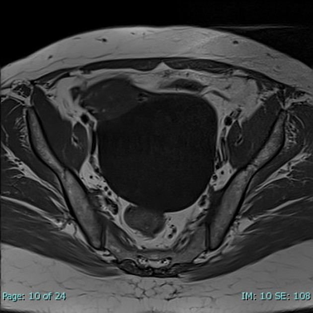
There is a large, well-defined, left adnexal cystic lesion mostly arising from the left ovary showing thin enhancing internal septation and the wall does not have mural nodules or papillary projections. It measures about 11.5 x 10.5 x 13 cm (AP, transverse and CC dimension).
There is also a right ovarian simple cystic lesion measuring about 2.4 x 3 cm.
There are numerous uterine fibroids , the largest appears subserosal and pedunculated , measuring about 6.9 x 5.5 cm , otherwise the uterus shows normal endometrial thickening and junctional zone .
Multiple Nabothian cysts are noted .
Vaginal septum is noted.
No significant free fluid seen.
No lesion in the urinary bladder .
Case Discussion
An adnexal tumor that manifests as a unilocular cystic mass with homogeneous signal intensity, a thin regular wall or septum, and no mural nodule or papillary projection, is compatible with a benign serous cystadenoma.


 Unable to process the form. Check for errors and try again.