
Traumatic intracranial hemorrhage with incidental carotid dolichoectasia mimicking sellar mass
Presentation
Sustained a fall, had a tonic clonic-seizure in the ER. Intracranial bleeding, fracture?
Patient Data


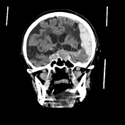
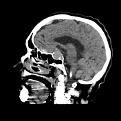
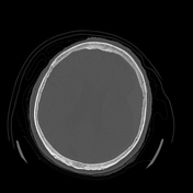
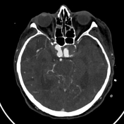
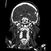
Large left frontal, parietal and temporal subdural hematoma.
A smaller subdural collection along the left tentorium is more subtle.
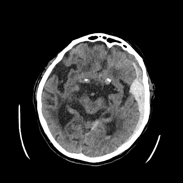
Left temporal pole contusion, with small subarachnoid bleeding also layering in the occipital horn of the lateral ventricles.
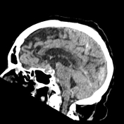
Prior righttemporal and left frontal extensive ischemic changes.
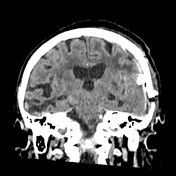
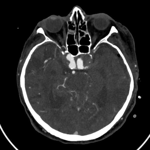
Incidental sellar expansile mass-like lesion showing suprasellar propagation (however the widening of the carotid canal contradicts this, and points towards vascular etiology). Stable compared to an earlier noncontrast CT (not shown). Further workup recommended.
Advanced (Fazekas III) chronic small vessel ischemic changes.


CTA a few days later after surgical drainage:
Staus post almost complete drainage of the left subdural hematoma. The collection along the tentorium is virtually unchanged.
Subarachnoid blood present in the ventricles and sulci at multiple locations bilaterally.
Bilateral carotid dolichoectasia, with circumferential fusiform aneurysms showing mural thrombi in the C4 segments, more pronounced on the left.
Significant atherosclerosis, about 50% narrowing of the intracranial right vertebral artery.
Incidental heterogeneous, partially cystic thyroid nodules.
Case Discussion
The case demonstrates multiple subdural, subarachnoid and parenchymal contusion hemorrhages with an incidental expansile sellar mass-like lesion, macroadenoma was suggested as a working diagnosis. However, subsequent CTA confirmed it as a result of bilateral extensive carotid dolichoectasia and aneurysms.
It is also a good example of reader fatigue and the perils of the satisfaction of search phenomenon since there is simultaneously a multitude of acute, neoplastic, and chronic pathologies present. The pituitary pseudolesion also illustrates anchoring bias.
Key take home message: we should train ourselves to increase, or at least maintain our alertness when we encounter significant pathology.


 Unable to process the form. Check for errors and try again.