
Adventitial bursitis secondary to a distal femoral osteochondroma
Diagnosis almost certain
Presentation
Medial knee pain aggravated by walking, of two weeks duration.
Patient Data
Age: 15 years
Gender: Male

Download
Info

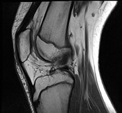
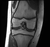
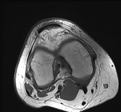
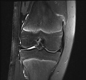
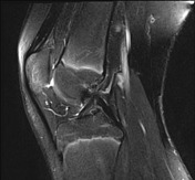
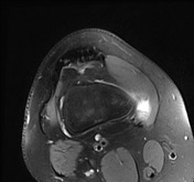
A sessile osteochondroma is noted arising from the medial aspect of distal femoral diaphysis measuring about 7 x 8 mm with overlying cartilage of about 1 mm in thickness. The lesion abuts the vastus medialis muscles.
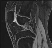
An isointense T1W and hyperintense T2W area are in the deep soft tissues underneath the vastus medialis muscle, suggestive of adventitious bursitis.
Case Discussion
This case beautifully illustrates adventitious bursa formation as a result of chronic soft tissue friction and irritation secondary to an osteochondroma.


 Unable to process the form. Check for errors and try again.