
Presentation
Abdominal pain and large suspicious focal lesion on the US.
Patient Data


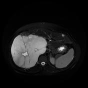
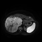
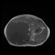
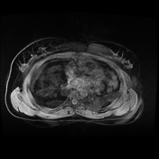
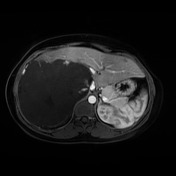
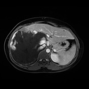

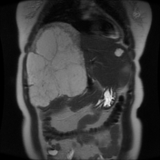
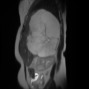
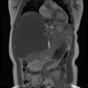

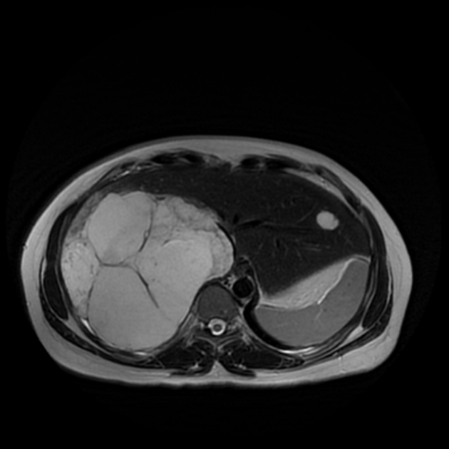
The right hepatic lobe is nearly totally replaced by a giant mass lesion measuring 16 x13 cm on its maximum axial dimensions and eliciting low signal on T1 WI and high signal on T2 WI showing cleft-like region of higher signal in keeping with a central scar. It shows peripheral nodular puddling of contrast material with progressive centripetal enhancement of the mass with a central area of low attenuation compatible with a fibrous scar. It exerts mass effect on the IVC which appears compressed.
Two small hepatic hemangiomas are seen on the left hepatic lobe with the same typical enhancement pattern.
Case Discussion
Giant hepatic hemangiomas are considered atypical hemangiomas, exceeding 5 cm in size.
Typical imaging features include early peripheral nodular puddling on initial post-contrast series with progressive centripetal enhancement on delayed series.
Giant hemangiomas might have central loculations or clefts of high signal intensity on T2; the high signal intensity is higher than that of the surrounding hemangioma parenchyma. These clefts are likely representing areas of cystic degeneration or liquefaction or central scar.


 Unable to process the form. Check for errors and try again.