
From the case:
Cavernous hemangioma - brain

Download
Info

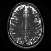
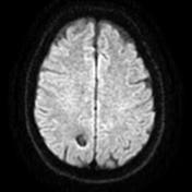
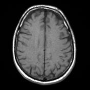
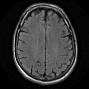
Selected axial images from an MRI demonstrate typical appearances of a cavernous hemangioma (cavernoma) with a circumscribed lesion with heterogeneous signal but profound signal loss and blooming on gradient echo. No evidence of surrounding edema to suggest a recent bleed.


 Unable to process the form. Check for errors and try again.