
Presentation
Gravida 0 parity 0, primary infertility for 2 years,
Patient Data
Age: 25 years
Gender: Female
From the case:
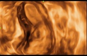
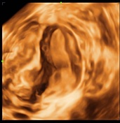
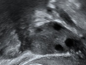
Unicornuate uterus (3D ultrasound)
Download
Info
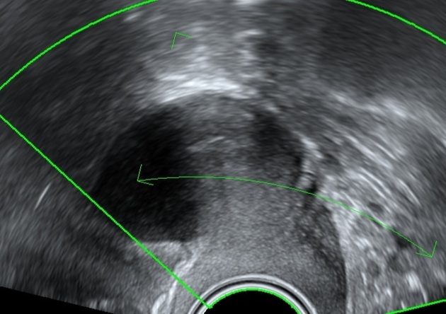
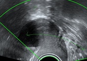
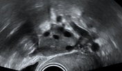
2D transvaginal ultrasound was done for this case showed normal sized ovaries with relative small sized uterus and normal cervix, the uterus was shifted to the right with displaced left ovary away from the uterus, 3D ultrasound was applied and showed unicornuate shaped uterus.
Case Discussion
Abnormal shape of the cavity is suspected when the sonographer finds difficulty in obtaining the mid-sagital view of uterus and cervix together in one view.


 Unable to process the form. Check for errors and try again.