
Presentation
Pelvic pain and palpable mass on physical exam.
Patient Data
Age: 65 years
Gender: Female
From the case:
Ovarian serous cystadenoma


Download
Info

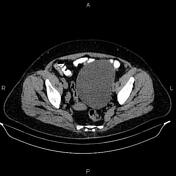
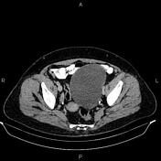
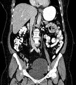
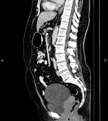
A 125×90 mm unilocular cystic lesion without enhancing solid components is noted in the left adnexa. The left ovary couldn’t be defined separately.
A few gas containing stones are noted in the gallbladder.
A 5 mm stone is seen in the lower calyx of the left kidney.
Case Discussion
Left adnexal cystic lesion; pathology proved ovarian serous cystadenoma, a type of benign ovarian epithelial tumor at the benign end of the spectrum of ovarian serous tumors.
On CT images, ovarian serous cystadenoma is often seen as a unilocular (typically) or multilocular cystic mass, with a thin regular wall or septum and no solid enhancing component.


 Unable to process the form. Check for errors and try again.