
Presentation
Not applicable
Patient Data
Age: 60 years
Gender: Male
From the case:
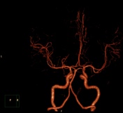
Trifurcated left middle cerebral artery


Download
Info
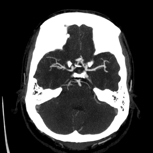
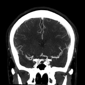
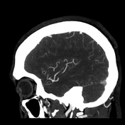
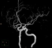
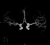
Cerebral CT angiography shows trifurcated M2 segment left middle cerebral artery.
Case Discussion
The commonest pattern (around 80%) of branching of the MCA is by bifurcation of the M2 segment into superior and inferior divisions. About 10% make trifurcations and another 10% split into more than three branches. One implication of such branching patterns is that the point of division is prone to aneurysms and pseudoaneurysms.


 Unable to process the form. Check for errors and try again.