Presentation
Work up for microscopic hematuria.
Patient Data
![](https://prod-images-static.radiopaedia.org/images/58449413/0._thumb.jpeg)
![](https://prod-images-static.radiopaedia.org/images/58449415/0._thumb.jpeg)
![](https://prod-images-static.radiopaedia.org/images/58449416/0._thumb.jpeg)
![](https://prod-images-static.radiopaedia.org/images/58449417/0._thumb.jpeg)
![](https://prod-images-static.radiopaedia.org/images/58449418/0._thumb.jpeg)
![](https://prod-images-static.radiopaedia.org/images/58449419/0._thumb.jpeg)
![](https://prod-images-static.radiopaedia.org/images/58449420/0._thumb.jpeg)
![](https://prod-images-static.radiopaedia.org/images/58449413/0._big_gallery.jpeg)
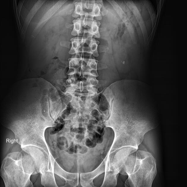
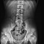
In non-contrast images, a small stone is noted in the lower part of the left renal lodge.
After contrast media administration, the enhancement of both kidneys is non-uniform. In the secretory phase, moderate hydronephrosis without hydroureter is evident bilaterally; the left is more severe than the right causing delayed contrast excretion.
![](https://prod-images-static.radiopaedia.org/images/58449453/32._thumb.jpeg)
![This study is a stack](/packs/stack-YQKLCKBI.gif)
![](https://prod-images-static.radiopaedia.org/images/58449515/33._thumb.jpeg)
![This study is a stack](/packs/stack-YQKLCKBI.gif)
![](https://prod-images-static.radiopaedia.org/images/58449586/46._thumb.jpeg)
![This study is a stack](/packs/stack-YQKLCKBI.gif)
![](https://prod-images-static.radiopaedia.org/images/58449453/32._big_gallery.jpeg)
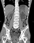
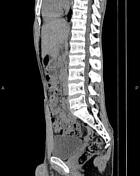
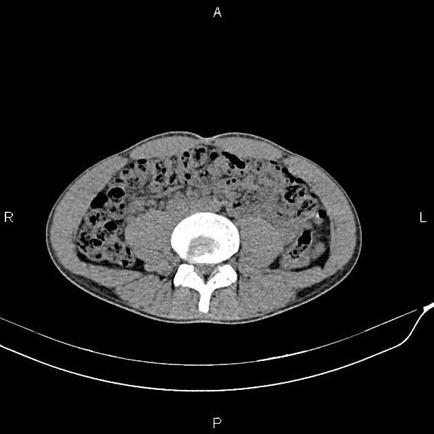
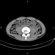
Moderate dilatation of the pelvicalyceal system is seen on both sides without ureter dilatation or ureteral stone.
A stone is also noted in the left kidney's lower part.
Case Discussion
Typical IVU and CT scan appearance of the bilateral ureteropelvic junction obstruction (UPJO).