
Presentation
Adult patient presenting with non-specific headache.
From the case:
Dural arteriovenous fistula

Download
Info
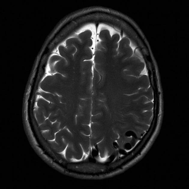
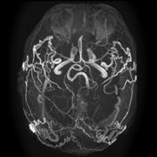
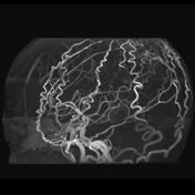
Large flow voids in the parietal region on the left are seen on this single T2 image which appear to be extra-axial. MIP MRA demonstrates very enlarge external carotid artery supply to a dural arteriovenous fistula.
From the case:
Dural arteriovenous fistula


Download
Info
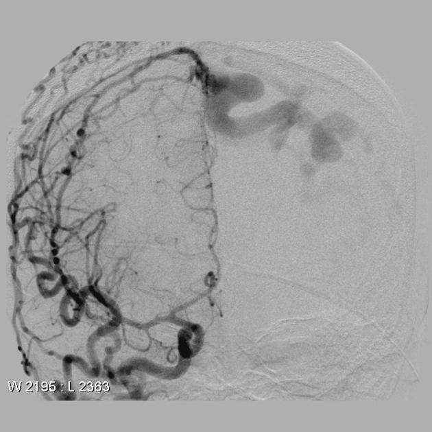
An angiogram was performed confirming the presence of dural arteriovenous fistula, located at the posterior aspect of the superior sagittal sinus, and draining via large cortical veins.
Case Discussion
This patient went on to have surgical ligation of a confirmed dural arteriovenous fistula.


 Unable to process the form. Check for errors and try again.