Presentation
History of brain tumor and surgery five years ago. Now the patient has been referred complaining of headaches and seizures.
Patient Data


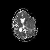
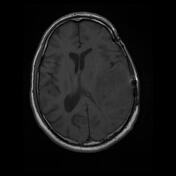

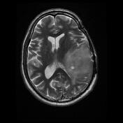
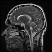
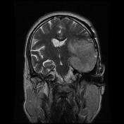
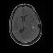
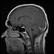
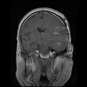
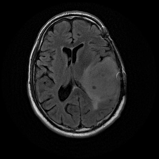
Evidence of prior craniotomy is present on the left side.
Large heterogeneous intra-axial mass measuring about 40 x 55 x 65 mm involving the left frontotemporal lobes grey and white matter that show a few internal cystic changes and punctate foci of enhancement.
There is a marked mass effect associated with vasogenic edematous changes, effacement of the left lateral ventricle occipital horn, uncal herniation, and midline shift to the right side measuring about 7 mm.
The mentioned mass shows water restriction on DWI images.
Few high signal foci in T2 and flair sequences at subcortical and periventricular white matter of both cerebral hemispheres depict microvascular ischemic events.

The patient went on to have surgery five years ago and the histopathological finding was compatible with oligodendroglioma, NOS type, and WHO grade II.
Case Discussion
Final diagnosis: recurrence of the known brain tumor (oligodendroglioma, NOS type, and WHO grade II).


 Unable to process the form. Check for errors and try again.