Presentation
Known chronic hepatitis C and liver cirrhosis.
Patient Data
Age: 60 years
Gender: Male
Note: This case has been tagged as "legacy" as it no longer meets image preparation and/or other case publication guidelines.
From the case:
Hepatocellular carcinoma


Download
Info

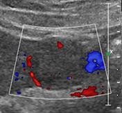
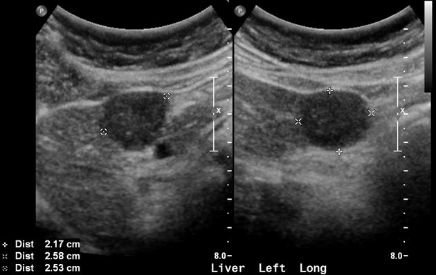
On surveillance US, a hypoechoic lesion was noted in segment 3 of the liver and demonstrated vascularity on Doppler study.
From the case:
Hepatocellular carcinoma


Download
Info

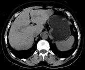
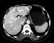
CT scan of the abdomen with quadriphasic liver protocol was performed which confirmed the presence of a hyper-vascular lesion in segment 3, demonstrating washout on portovenous phase.
Case Discussion
A diagnosis of hepatocellular carcinoma was entertained.


 Unable to process the form. Check for errors and try again.