
Presentation
Diplopia on lateral gaze
Patient Data
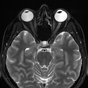

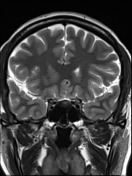

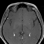
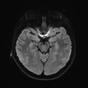
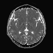
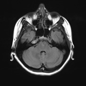
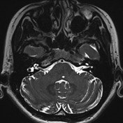
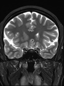
Ovoid T2 hyperintense lesion in the posterior aspect of the pons just to the left of midline measuring 3 mm. The lesion is in the expected region of the left abducens nucleus. No associated contrast enhancement, diffusion restriction or expansion. The cisternal portion of the left abducens nerve is smaller than the right, but appears intact.
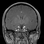
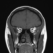
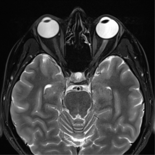
Medial deviation of the left globe. The left lateral rectus demonstrates moderate atrophy. The remainder of the extra-ocular muscles have a normal appearance.
Conclusion: Stable appearance of a longstanding pontine T2 hyperintense lesion in the region of the left abducens nucleus. This is most likely the cause of the left abducens nerve paresis, evidenced by medial gaze deviation and lateral rectus atrophy. Smaller left abducens nerve compared to the right presumably represents atrophy secondary to the involved abducens nucleus.


 Unable to process the form. Check for errors and try again.