
Presentation
Acute presentation, non-productive cough, pleuritic chest pain on left side and shortness of breath
Patient Data
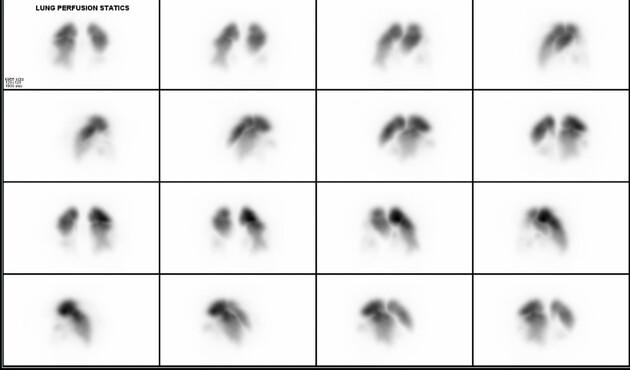
MIP SPECT images demonstrate large perfusion defects bilaterally affecting the lower lobes predominantly

Axial SPECT images demonstrate large perfusion defects bilaterally affecting the lower lobes predominantly.
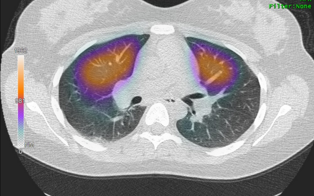
Fused SPECT-CT images demonstrate large perfusion defects bilaterally affecting the lower lobes predominantly, which are not matched on the CT component of the study.
The CT component demonstrates a small amount of peripheral ground-glass change in the posterior right lung lower lobe, which may represent developing infarction.
Appearances are in keeping with large volume bilateral pulmonary emboli.
Case Discussion
During the COVID pandemic, the ventilation portion of a VQ scan was often not performed as it was deemed an aerosol generating procedure and therefore a potential risk to the nuclear medicine staff.
The CT chest, as in this example, was utilized as a surrogate for the ventilation portion of the study.


 Unable to process the form. Check for errors and try again.