
Presentation
Pain in the right iliac fossa for the last 10 days. No fever or vomiting.
Patient Data

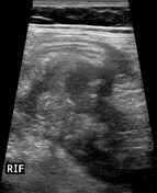
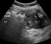
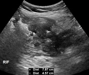
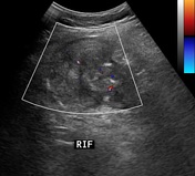
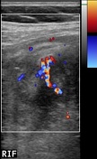
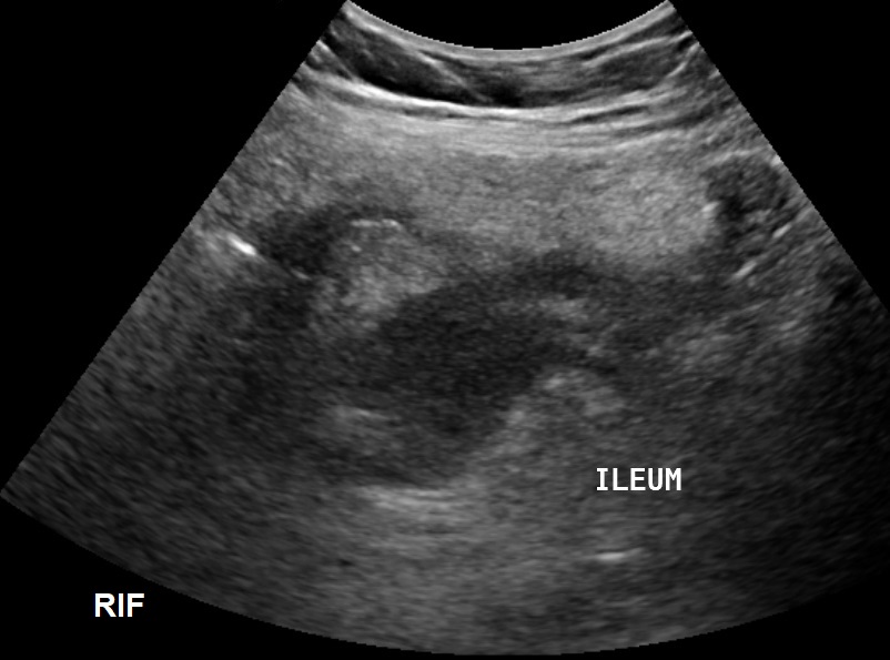
large irregular heterogeneous echogenicity lesion in the right iliac fossa, in relation to the thick-walled cecum and terminal ileum measuring approximately 79 x 46 mm. The lesion has mild internal vascularity on color Doppler ultrasound examination
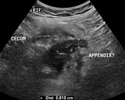
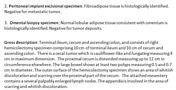
tubular blind-ended non-compressible structure measuring 8 mm seen along its medial aspect, which is likely the appendix
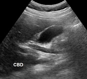
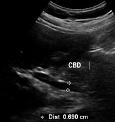
prominent CBD measuring 7 mm without any obvious filling defect


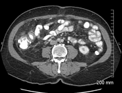
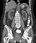
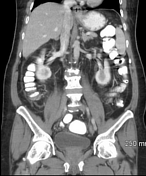
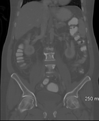
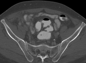
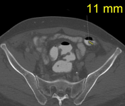
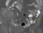
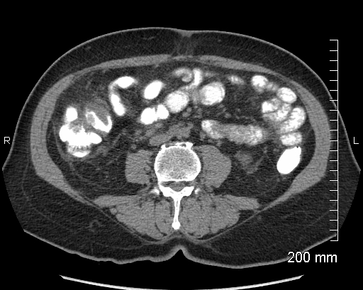
irregular mural thickening of the terminal ileum and cecum associated with an irregular heterogeneously enhancing mass lesion surrounded by fat stranding and locoregional lymphadenopathy with the largest lymph node measuring 14 mm in its transverse diameter, no evidence of distant metastases
the appendix could not be separately identified from the mass and no signs of intestinal obstruction
small pedunculated filling defect, likely a polyp, seen in the distal descending colon
prominent CBD measuring 7 mm (likely age-related)
small splenule measuring 9 mm, seen adjacent to the anterior aspect of the main spleen.
transitional vertebra at the lumbosacral junction (sacralized 5th lumbar vertebra). Degenerative changes in the visualized spine
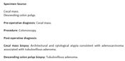
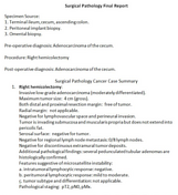
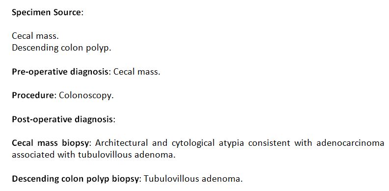
Histopathology reports of the colonoscopic biopsy of the cecal mass and colonic polyp revealed an adenocarcinoma of the cecum and a tubulovillous adenoma.
Case Discussion
Taking into consideration the age of the patient, CT scan features are suspicious for a neoplastic process, like cecal carcinoma as histologically proven in this case. Another possible differential would be lymphoma. The possibility of an appendicular mass or ileocecal tuberculosis is less likely in this case.


 Unable to process the form. Check for errors and try again.