
Presentation
Chronic abdominal pain and elevated CA19-9.
Patient Data


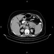
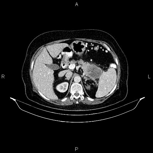
A large, ill-defined mass with progressive ring enhancement and areas of central necrosis is present in the pancreatic tail. The mass encases the spleen vessels and abuts the adjacent spleen and left adrenal gland. Several dilated collateral vessels are seen around the splenic hilum. Mild surrounding fat stranding is evident.
Several gas-containing stones are seen in the gallbladder.

Regarding imaging findings and loss of metastasis and criteria for resectability, the patient underwent distal pancreatectomy, splenectomy and simultaneous cholecystectomy. The histopathology evaluation and IHC findings confirmed a well-differentiated adenosquamous carcinoma of the pancreas, a rare pancreatic malignancy.
Case Discussion
On CT images, adenosquamous carcinoma of the pancreas appears as an ill-defined, lobulated hypo or iso-attenuating mass. Most tumors show progressive ring enhancement and central necrosis, differentiating them from pancreatic ductal adenocarcinoma. Calcification and hemorrhage are not typical features of these tumors.


 Unable to process the form. Check for errors and try again.