
Presentation
55-year-old female with decreased range of motion and left shoulder pain increasing over the last 2 months.
Patient Data
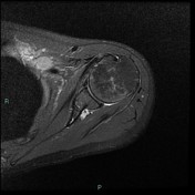

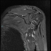
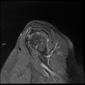
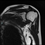
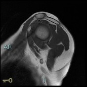

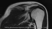
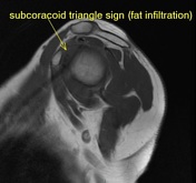
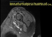
Sagittal T1-weighted sequences show the complete obliteration of subcoracoid fat triangle and distinct fatty tissue surrounding the coracohumeral ligament as having disappeared, and the coracohumeral ligament cannot be measured. The coronal T2-weighted fat-suppressed image demonstrates an abnormally thickened inferior glenohumeral ligament and the axillary pouch is contracted and poorly distended. There is also supraspinatus tendinopathy without tear.

Annotated images of adhesive capsulitis.
Case Discussion
Adhesive capsulitis is a self-limited clinical syndrome characterized by painful, gradual loss of active and passive glenohumeral motion. Replacement of the fat in the rotator interval with the abnormal soft tissue that can encase the biceps anchor (subcoracoid triangle sign), can be classified on a sagittal T1-weighted image as (A) absent, (B) partial, (C) complete.


 Unable to process the form. Check for errors and try again.