
Presentation
Chronic abdominal pain. GP-requested CT abdomen demonstrated adrenal lesions. The patient was referred to hospital for further evaluation. No history of primary malignancy. No infectious symptoms.
Patient Data


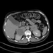
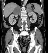
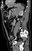
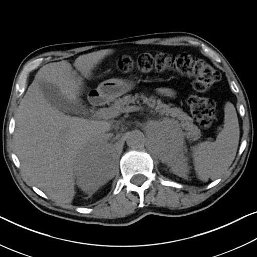
Large bilateral soft-tissue density adrenal lesions. No internal fat or calcification.
LEFT ADRENAL LESION
Pre-contrast: 33 HU
Portal venous contrast: 59 HU
15 minute delayed: 58 HU
Absolute washout: 3.8%
Relative washout: 1.7%
RIGHT ADRENAL LESION
Pre-contrast: 28 HU
Portal venous contrast: 33 HU
15 minute delayed: 30 HU
Absolute washout: 60%
Relative washout: 9.1%
The washout characteristics for the left adrenal lesion are indeterminate. The right adrenal lesion is not enhancing, which is more characteristic of a cyst or haematoma.
Size >4cm is concerning for malignancy, including metastases or bilateral adrenal cortical carcinomas, although the lack of enhancement on the right does not suggest carcinoma.
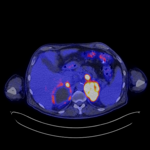
FDG-avid left adrenal lesion with numerous malignant FDG avid lymph nodes surrounding both adrenal glands near the upper abdominal aorta and IVC. No evidence of metastatic disease elsewhere.
The most likely aetiology was considered bilateral adrenal metastases with metastatic lymph nodes in the upper abdomen, but a primary malignancy was not identified.
Synchronous primary adrenal carcinomas with adjacent metastatic nodes was considered possible.
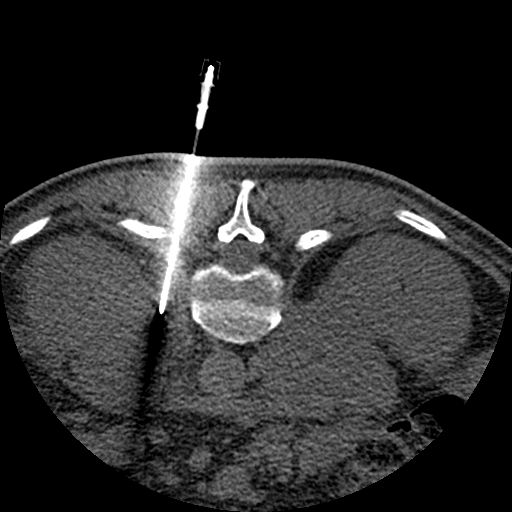
Left adrenal biopsy: High grade B cell lymphoma, consistent with diffuse large
B cell lymphoma (DLBCL), non-germinal centre B (GCB) subtype.
Case Discussion
Following the histology, bone marrow aspiration was performed which was also consistent with DLBCL.
Primary adrenal lymphoma is a rare entity. Secondary involvement of the adrenal glands is more common.


 Unable to process the form. Check for errors and try again.