
Presentation
Abdominal pain and an adrenal mass on ultrasound exam.
Patient Data
Age: 50 years
Gender: Female
From the case:
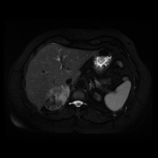
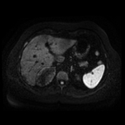
Adrenal myelolipoma - MRI


Download
Info
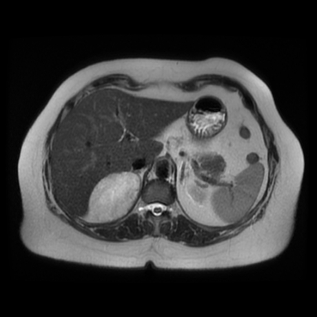
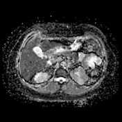
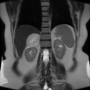
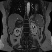
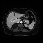
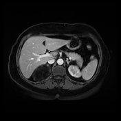
Right adrenal large well-defined soft tissue mass lesion eliciting high signal on T2 WI with partial signal suppression on fat saturation and out-of phase sequences. No diffusion restriction or postcontrast enhancement of the mass.
From the case:
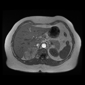
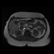
Adrenal myelolipoma - MRI
Download
Info
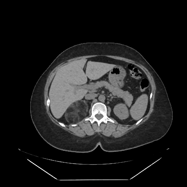
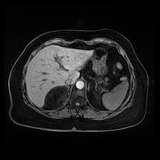
Right adrenal large mass of predominant macroscopic fat-density, confirming the diagnosis of adrenal myelolipoma.
Case Discussion
CT appearance of adrenal myelolipoma is usually characteristic of fat-containing lesion, however, MRI features may be somewhat confusing.
MRI features of adrenal myelolipoma include:
- T1: typically hyperintense due to fat contents
- T1 (FS): shows fat suppression
- T2: generally intermediate to hyperintense but can sometimes vary depending on contents (especially blood products)
- T1 C+ (Gd): shows striking enhancement 1
- in and out of phase: in masses with mixed components, out of phase imaging may demonstrate signal loss as the microscopic fat cells usually have little intracellular water


 Unable to process the form. Check for errors and try again.