
Presentation
Suspicious ascites in a young woman. ? Carcinomatosis due to ovary cancer
Patient Data


Unusual findings for a supposed ovarian cancer.
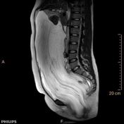
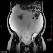
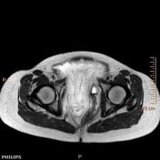
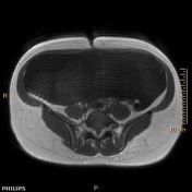
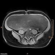
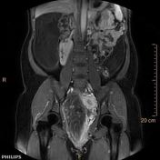
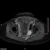
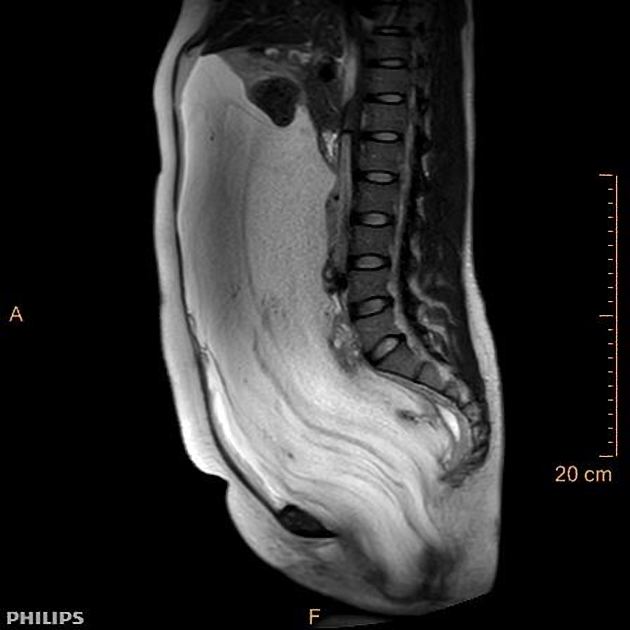
Actually the main lesion originates from the subperitoneal space: it most likely comes from the perineum, breaks through the puborectal and levator ani muscles, extends into the right ischiorectal fossa and pushes to the left the rectum, uterus and bladder, without invading them. It then most likely extends in the retroperitoneal space up to the upper abdomen, behind the liver. The right colon is pushed away to the left, actually the entire digestive tract is pushed away as a whole.
The lesion features mostly a liquid-like content, probably myxoid, (NOT ascites), T2 hyperintense, T1 hypointense with small T1FS hyperintense spots (blood or proteins), without fat.
Interestingly there are long thin "filaments" enhancing after contrast injection. These are oriented vertically in a layered "onion-peel"-like fashion.
Case Discussion
Careful examination of the MRI revealed a primarily cystic tumor arising from the subperitoneal space, with most likely a myxoid main component. Uterus and ovaries were normal.
Using a gamuts 1 for pelvic extraperitoneal masses, possible diagnoses for a cystic mass with myxoid stroma include myxoma, aggressive angiomyxoma, schwannoma, plexiform neurofibroma.
Judging by the young age of the female patient, the subperitoneal location with puborectal and levator ani muscles rupture, the myxoid component associated with an enhancing vascular component in layered and "onion-peel"-like appearance, our most likely diagnosis was aggressive angiomyxoma 2.


 Unable to process the form. Check for errors and try again.