
Presentation
Withheld
Patient Data
Age: 50 years
Gender: Male
From the case:
Alpha-1-antitrypsin deficiency


Download
Info
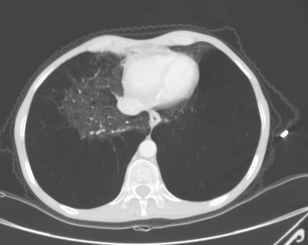
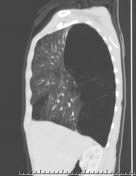
Panlobular emphysematous changes diffusely replacing the lower lobes of both lungs with very thin internal septa but no residual lung tissue seen. Similar, yet, less severe changes are seen in the medial lobe of the right lung where interspersed lung tissue is seen.
Centrilobular and paraseptal emphysematous changes involving the upper lobes of both lungs.
Subsegmental area of consolidation with air bronchograms seen in the inferior segment of the lingula.
Case Discussion
Severe lower lobe predominant panlobular emphysematous changes with no residual lung tissue seen. In a middle aged patient, these changes may be secondary to alpha-1-antitrypsin deficiency.


 Unable to process the form. Check for errors and try again.