
Presentation
History of increasing hip and lower back pain for years.
Patient Data


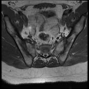
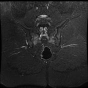
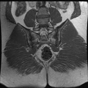
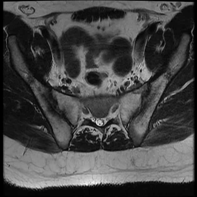
Both sacroiliac joints show more or less symmetrical subchondral marrow edema signal being of high T2 / STIR and low T1 signal mixed with some sclerotic signal which is low in all sequences. These changes are more marked at the sacral aspect of both articulations.
Similar subchondral marrow edema signal is noted at the anterior and posterior upper corners of L4 and lower corners of L3 vertebral bodies with normal height and bright T2 signal of L3/L4 disc.
Case Discussion
Bilateral rather symmetrical sacroiliitis. In light of the patient’s age and gender, the above-described changes are suggestive of ankylosing spondylitis, for further clinical and laboratory correlation with HLA-B27.


 Unable to process the form. Check for errors and try again.