
Presentation
Intermittent rectal bleeding.
Patient Data

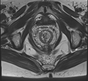
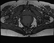
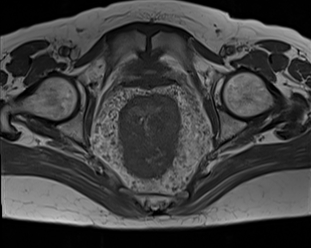
There is a regular circumferential thickening of the anorectal wall with numerous calcifications (phleboliths) within and in perirectal space. Multiple perirectal serpiginous structures, suggestive of vascular origin.


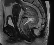
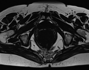
The MRI sequences demonstrate:
- marked and regular circumferential mucosal and submucosal thickening of the anorectal region, 12 cm of length from the anal margin.
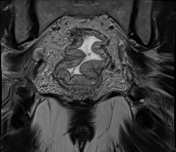
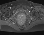
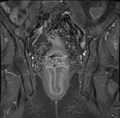
- numerous enhancing serpiginous vessels within the anorectal wall well-visualized on postcontrast fat sequences
- increased signal within the perirectal fat with numerous serpiginous flow void structures with enhancement on postcontrast sequences (small vessels supplying the hemangioma/vascular malformation)
- small punctuate phleboliths of low signal within the anorectal wall as well as in the perirectal space
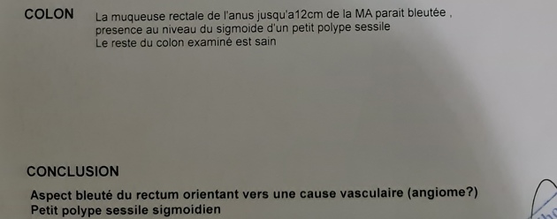
Colon
- the rectal lining of the anus up to 12 cm from the anal margin appears bluish
- small sessile polyp of the sigmoid colon
- the rest of the colon was normal
Conclusion
- bluish appearance of the rectum pointing to a vascular cause (hemangioma?)
- small polyp of the sigmoid colon
Case Discussion
The clinical presentation, colonoscopic findings, and the CT/MRI features suggest an anorectal hemangioma/vascular malformation.
On imaging the main differential diagnosis is the hemorrhoids which can give the same appearance on T2, however, the lack of extension to the perirectal fat exclude this possibility 1.
Hemangiomas and vascular malformations are considered as rare entities 1,2. They may involve any intestinal segment, nevertheless, the small bowel is the most frequently affected 1. Anorectal and colonic involvement is rarer 1.
Additional contributor: A. Ramdani, MD


 Unable to process the form. Check for errors and try again.