
Presentation
Withheld.
Patient Data
Age: 18 years
Gender: Male


Download
Info
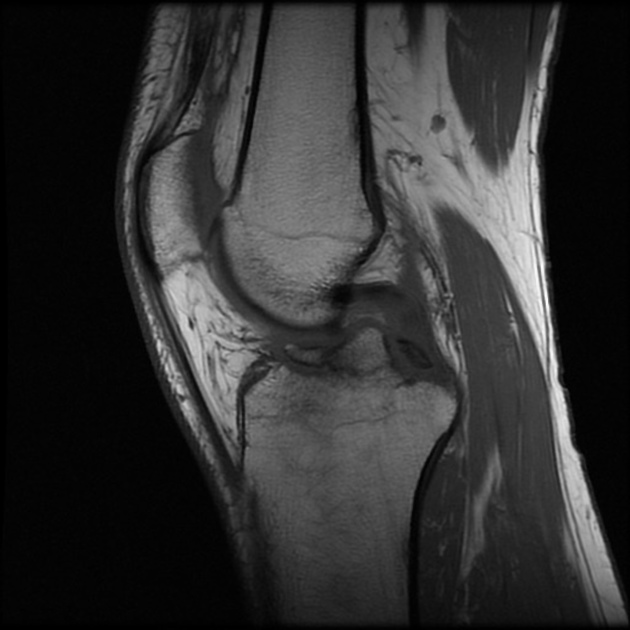
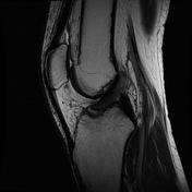
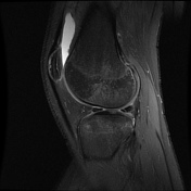
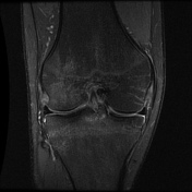
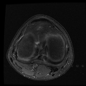
- anterior tibial spine fracture with 2mm fracture line gap
- extensive marrow edema in lateral tibiofemoral condyles
- vertical oblique tear of posterior horn of lateral meniscus
- mild flattening of the ACL fibers which appears intact
Case Discussion
The anterior tibial spine fracture was evident in the radiograph itself. The knee MRI was done in this case to assess if there is any associated internal derangement,


 Unable to process the form. Check for errors and try again.