
Presentation
Blocked right nasal passage, with sinus symptoms and chronic headaches.
Patient Data
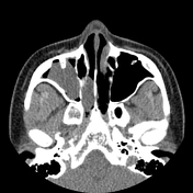

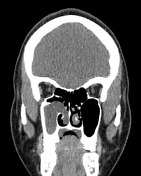

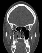

Non-contrast CT imaging demonstrates soft tissue attenuation of the right maxillary antrum, with the widening of the maxillary ostium and osteomeatal complex. There is medial and posterior extension into the nasal cavity. The mass terminates within the nasopharynx. There is no bone erosion evident, with minimal maxillary antral bony remodeling.
There is bilateral concha bullosa. There are bilateral Onodi (sphenoethmoidal) air cells.
Case Discussion
Features suggestive of a right-sided antrochoanal polyp with far posterior extension past the posterior choana to appear as a hanging mass within the nasopharynx that is identified on clinical oral cavity visualization. The mass measured 10-20 Hu with the noted absence of any calcification or dense components.
Concha bullosa and especially Onodi air cells are important normal variants to mention in preoperative CT assessments for FESS.


 Unable to process the form. Check for errors and try again.