
Presentation
Mild intermittent abdominal pain in the right iliac fossa. Normal WBC count.
Patient Data
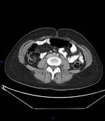

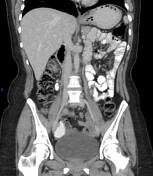
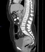
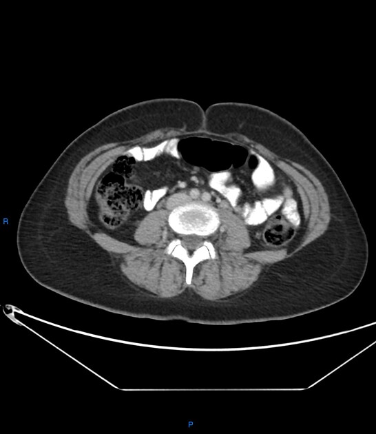
The appendix is thickened, measures up to 15-20 mm, with a minimal blurring of the surrounding fat planes yet no masses or collections.
Bulky uterus shows few junctional zone cystic changes, left adnexal cystic lesion.
The patient was subjected to an elective laparoscopic appendectomy.
The histopathological report
SPECIMEN:
Laparoscopic appendectomy
MACROSCOPY:
Received in a labeled container with patient identification and fixed in formalin; consists of an appendix measuring 4.5 cm long & 3 cm in diameter. The cut section revealed a wall thickness; of 1 cm. Representative sections were taken in 4 cassettes.
MICROSCOPY:
Sections examined revealed appendix entangling bland-looking endometrial glands, surrounded by endometrial stroma.
DIAGNOSIS:
Laparoscopic appendectomy:
Picture consistent with '' Endometriosis ''.
No evidence of dysplasia or malignancy.
Case Discussion
The clinical manifestations of the patient were not definite for acute appendicitis. The CT picture directs toward elective laparoscopic appendectomy. The histopathology surprisingly revealed appendical endometriosis.
The gastrointestinal tract is involved in endometriosis in 12-37% of patients.
Appendiceal endometriosis should be considered in the differential diagnosis of women of reproductive age complaining of right iliac fossa pain especially if the clinical, laboratory, and imaging features are not typical for acute appendicitis.
The presence of other imaging features of endometriosis (adenomyosis, adnexal lesions) as seen in this case, can help in this suspicion on imaging.


 Unable to process the form. Check for errors and try again.