
Presentation
Abdominal pain.
Patient Data
Age: 65 years
Gender: Female
From the case:
Appendiceal mucocele


Download
Info
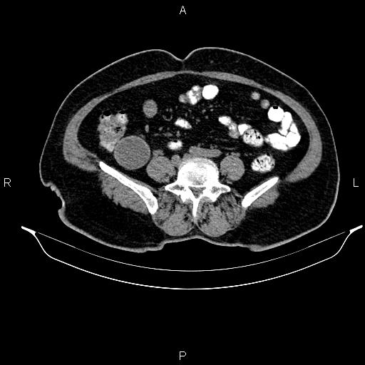
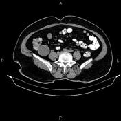
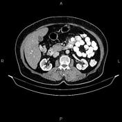
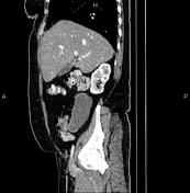
A 105 × 50 × 47 mm well-circumscribed tubular, elongated cystic lesion with fine incomplete peripheral calcification is seen in the right iliac fossa extends to the subhepatic space. The lesion is lying vertically and is continuous with terminal end of the appendix. No surrounding fat stranding or enlarged lymph node are evident.
The hepatic attenuation value is less than of the spleen, suggesting fatty liver. A few small calcified foci are seen at liver parenchyma most consistent with healed granuloma.
A few tiny stones are seen at both kidneys less than 3 mm.
Degenerative changes as osteophytosis are seen at the lumbar spine.
Case Discussion
Features are typical for appendiceal mucocele.


 Unable to process the form. Check for errors and try again.