
Presentation
Right flank pain that began 3 days before presenting to the ER, that wandered to the right lower abdomen .
Patient Data


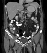

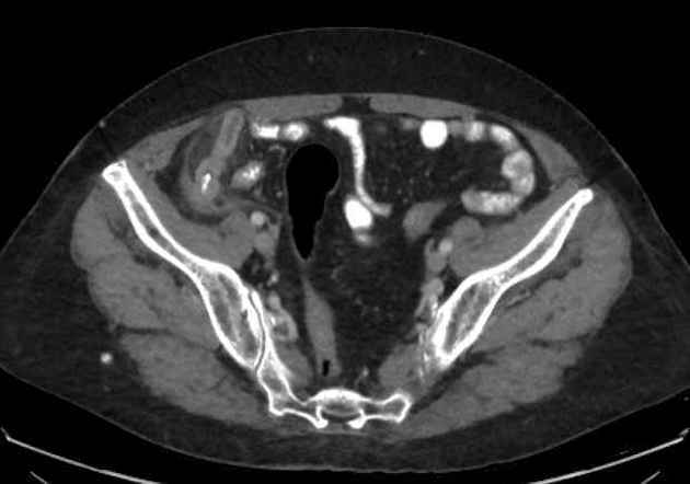
The appendix is thickened, measuring up to 14 mm in diameter, hyperemic, with periappendiceal fat stranding. There is a sharp bone (probably a chicken fibula) in its proximal lumen that is seen perforating its wall near its origin.
2.4 cm subcapsular cyst in segment VI of the liver.
Spondylotic and discogenic degenerative changes in the spine with reversal of the lumbar lordosis.
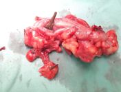
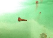
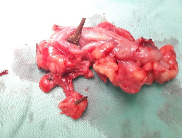
A chicken's fibula was revealed in the removed appendix.
Case Discussion
The swallowed chicken fibula became lodged in the appendix, leading with its blunt tip. The sharp tip of the broken fibula (its anatomical tip is even sharper) can be seen outside the appendiceal wall.
The radiologist warned the surgeons in advance to take care not to be pricked by the tip of the bone and was given extra credit post-surgery for having identified the avian bone on CT.
Interestingly, when a pin or sewing needle is swallowed, the leading tip is usually the blunt one (also from personal experience of having reported dozens of such cases).


 Unable to process the form. Check for errors and try again.