
Presentation
3 years of mild bifrontal headache.
Patient Data
Age: 15 years
Gender: Male
From the case:
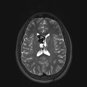
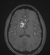
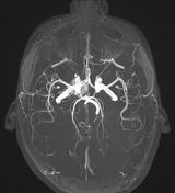
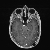
Arteriovenous malformation in the lateral ventricle


Download
Info

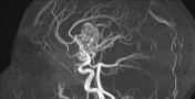
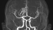
At the supratentorial level in the right caudate nucleus, multiple abnormal vessels are consistent with an arteriovenous malformation. It is supplied by the right middle cerebral and anterior cerebral arteries. Venous drainage is primarily to the internal cerebral vein and subsequently to the great cerebral vein. The lesion extends to the interior of the frontal horn of the right lateral ventricle with partial involvement of the internal capsule.
Left parietooccipital VP shunt.
Case Discussion
Although arteriovenous malformations are usually within the substance of the brain, when subependymal, they can bulge into the ventricle as is the case in this instance.


 Unable to process the form. Check for errors and try again.