
Presentation
G6P5 at 21 weeks gestation. Routine ultrasound.
Patient Data
Age: 40 years
Gender: Female
From the case:
Autosomal recessive polycystic kidney disease - antenatal


Download
Info
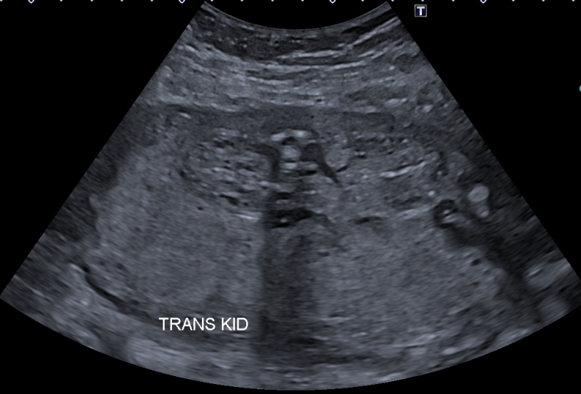
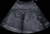
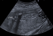
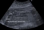
Echogenic enlarged kidneys with loss of the corticomedullary differentiation, and preserved reniform shape, containing numerous tiny cysts (size < 5mm) with associated severe oligohydramnios. The urinary bladder is not visualized, indicating most likely a lethal form of autosomal recessive polycystic kidney disease.
Case Discussion
Ultrasound findings of an autosomal recessive polycystic kidney disease (ARPKD) with severe oligohydramnios.


 Unable to process the form. Check for errors and try again.