
Presentation
Cough. Rule out pneumonia.
Patient Data
Age: 25
Gender: Female
From the case:
Azygos lobe

Download
Info

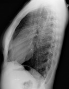
Azygos lobe. Otherwise, the chest x-ray is normal, without signs of pneumonia.
Case Discussion
An azygos lobe, while not a true accessory lobe, is a common anatomical variant. It is demonstrated in the right upper zone of frontal chest x-rays and presents as a linear density coursing towards the right hilum with a typical 'teardrop' at its inferior aspect representing the azygos vein.


 Unable to process the form. Check for errors and try again.