Presentation
History of a stroke three years ago referred with recurrent aspiration and pneumonia.
Patient Data

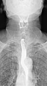

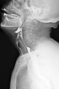
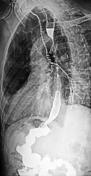
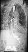
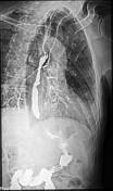
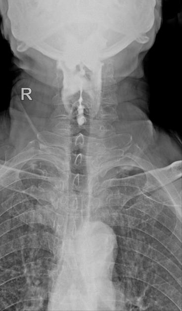
Lateral and oblique images show pooling of contrast in the pharynx with the aspiration of the barium contrast into the airway and inside the bronchial tree.
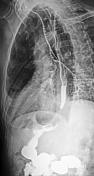
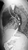
Evidence of oesophagal dysmotility also is seen.
The prominent aortic arc with pressure effect on the thoracic oesophagal barium column is noted.
Thoracic vertebral degenerative changes as endplate sclerosis and marginal osteophyte formation also are seen.
Case Discussion
The fluoroscopic study is the imaging modality of choice for the investigation of dysphagia.
It represents a good non-invasive, rapid and cost-effective diagnostic exam, and can show esophageal morphology, distensibility, and contractility.


 Unable to process the form. Check for errors and try again.