
Presentation
Found unresponsive.
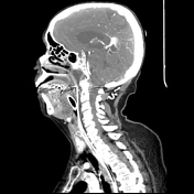
Patient Data


There is an ill-defined hypodensity within the ventral pons.
Qualitative analysis of cerebral perfusion maps demonstrate the proximal cerebral blood flow and cerebral blood volume within the central pons. There is also elevation of mean transit time and time to peak within similar locations.
There is occlusion of the mid basilar artery distal to the origin of the anterior inferior cerebellar arteries and proximal to the origins of the superior cerebellar arteries. There is distal reconstitution at the basilar apex with normal enhancement of the superior cerebellar and posterior cerebral arteries bilaterally.


AP and lateral cranial views were obtained which demonstrated a mid basilar occlusion.
A thrombectomy was performed (second series). AP and lateral cranial views revealed recanalization of the basilar artery.
Case Discussion
This is a case of an occlusion of the basilar artery.
The patient's treatment course was complicated by subarachnoid hemorrhage secondary to the angiographic procedure. This led to obstructive hydrocephalus for which in extraventricular drain was placed for several days. Eventually, the patient was discharged to a rehabilitation facility.
Co-author:
Trevina Soliman


 Unable to process the form. Check for errors and try again.