
Presentation
Decreased GCS, vomiting and right facial droop.
Patient Data
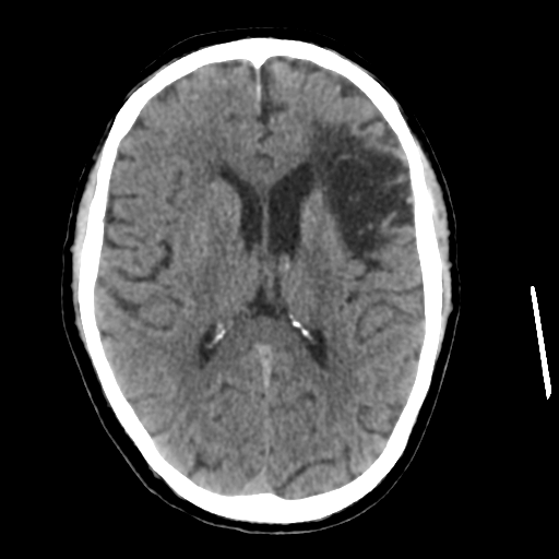
Old left MCA territory infarct with encephalomalacia and gliosis. Elsewhere grey-white differentiation is preserved with no hyperdense vessel seen. No intracranial hemorrhage.


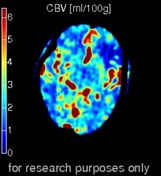
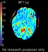
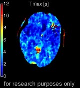
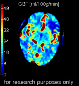
Marked increase in MTT / Tmax in the posterior circulation in particular the cerebellum and brainstem. CBF and CBV are only mildly reduced. Suggests changes largely of ischemic penumbra rather than a large infarct.
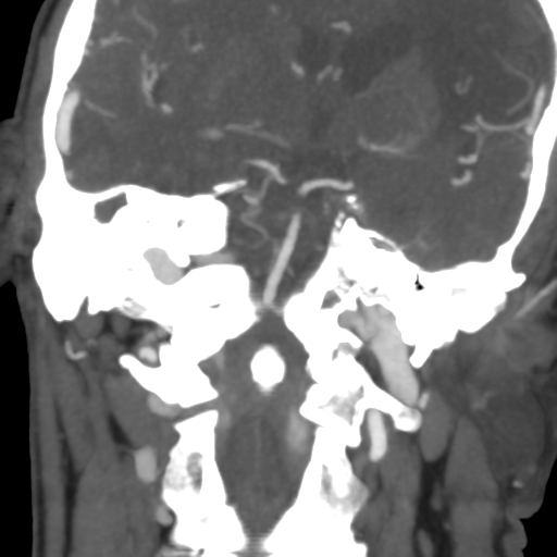
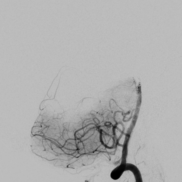
Filling defect within the distal basilar artery. Supply to the PCAs via PCOMs bilaterally.

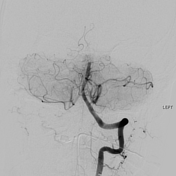
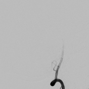
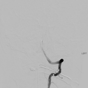
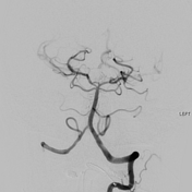
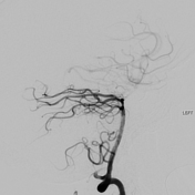
Initial left vertebral artery demonstrates filling defect in the distal basilar artery with no flow into the posterior cerebral arteries. Clot retrieval (mechanical thrombectomy) was successful with complete recanalization of the basilar artery and both posterior cerebral arteries.
Case Discussion
This is a case of don't be fooled by the initial non-contrast CT. There is an old stroke but nothing to explain the patient's new sudden onset symptoms. Perfusion clearly shows a large area of perfusion abnormality in the posterior circulation with CTA and DSA both showing a filling defect.
Clot retrieval (mechanical thrombectomy) has become standard of care for patients with some acute stroke patients. Important factors in patient selection includes relatively proximal thrombus, severity of symptoms, and lack of large established infarct.


 Unable to process the form. Check for errors and try again.