
Patient Data


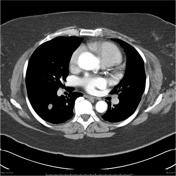
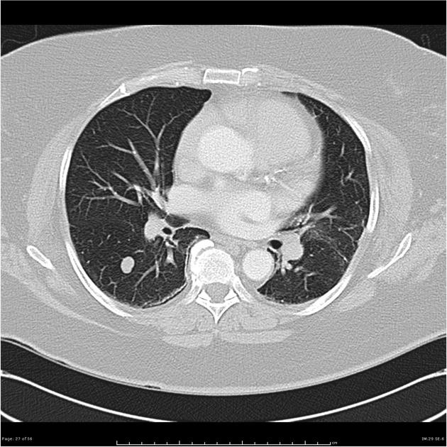
There are multiple discrete pulmonary nodules within both lungs, which are unchanged in number and size from the previous CT. The largest is a 13mm nodule in the superior segment of the RLL.
Case Discussion
VATS biopsy of a left lower lung nodule showed:
"Sections of the tumour mass show it to consist of bundles of smooth muscle fibres surrounded by a fibrous tissue stroma. Amongst the smooth muscle and fibrous tissue, there are a number of entrapped glandular structures lined by cuboidal to columnar epithelium. These are more prominent at the periphery of the lesion. The tumour cells show positive staining with smooth muscle actin, desmin and also with oestrogen and progesterone receptors. The entrapped glandular tissue shows positive staining with TTF-1. The features, including positive staining with hormone receptors, are typical of benign metastasising leiomyoma."
This case is just an example of an interesting pathology - there are no specific imaging features that would suggest metastasising leiomyoma.


 Unable to process the form. Check for errors and try again.