
Presentation
Primary infertility and history of pelvic inflammatory disease.
Patient Data
Age: 40 years
Gender: Female
From the case:
Bilateral tubal block and hydrosalpinx

Show annotations
Download
Info
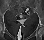
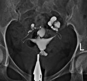
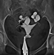
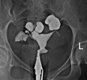
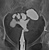
The body of the uterus is situated in the midline of the pelvic cavity and has a normal shape and contour.
The cervical canal is of normal length and is normally expanded.
The fallopian tubes are gradually filled and demonstrate abnormal length, diameter, and peristalsis, with retained contrast material and minimal spillage due to bilateral hydrosalpinx.
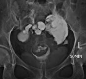
In the delayed imaging, contrast material is still observed in the fallopian tubes and the peritubal spaces, suggesting the presence of pelvic adhesions.
Case Discussion
Hysterosalpingography findings were compatible with bilateral tubal block and hydrosalpinx because of peritubal adhesions.


 Unable to process the form. Check for errors and try again.