
Presentation
Mild right hypochondrial pain over 2 months
Patient Data
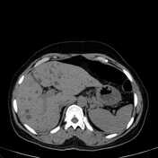

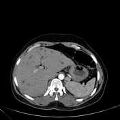
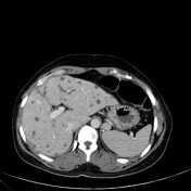
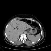
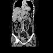

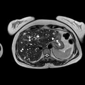
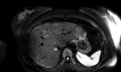
Mildly enlarged liver with multiple well-defined small-sized hypoattenuating non-enhancing focal lesions representing cysts. They are scattered in both liver lobes, the largest measuring 2.2 cm in diameter seen at segment VII of the right lobe.
Cervical enlargement with ill-defined thickening of the cervical canal.
Left simple cortical renal cyst, measures 1.6 x 2 cm.
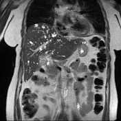

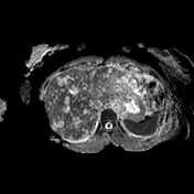

MRCP shows multiple well-defined small non-communicating cystic lesions scattered at both liver lobes with no definite communication with the biliary tree. No detected biliary dilatation.
They display very high signals on T2 images with free diffusion.
A hepatic biopsy was performed and revealed:
Microscopic examination: an examination of prepared slides revealed multiple cores of liver tissue that show large irregular fibromuscular spaces, lined by flat endothelial cells & devoid of RBCs.
No detected atypia or malignancy in examined material.
Final diagnosis: biliary microhamartomas.
Courtesy of DR. Nagwa Mokhtar pathology department, Mansoura faculty of medicine.
Case Discussion
Radiological examination supports the diagnosis of multiple cystic hepatic lesions. In favour of multiple biliary hamartomas rather than metastatic process.
A punch biopsy from the cervix revealed chronic non-specific cervicitis.
The patient was referred to a pain clinic and is now under regular follow up.


 Unable to process the form. Check for errors and try again.