
Presentation
Work up for chest pain.
Patient Data


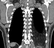
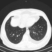

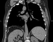
Fat-density tissue is uniformly seen in the lower posterior part of the left hemithorax. The distribution and placement of fat tissue at the base of the left hemithorax are more reminiscent of extrapleural fat.
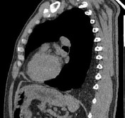
However, a focal defect is apparent at the posterior aspect of the left hemidiaphragm, and some omental fats are herniated through it.
The patient was called for a CT scan in the prone position to differentiate extrapleural fat from fat herniation through the diaphragmatic defect (Bochdalek hernia).
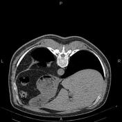
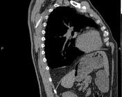

The CT images in the prone position show the fat tissue at the base of the left hemithorax is displaced anteriorly, suggesting herniated fat.
Case Discussion
Extrapleural fat is a benign condition and refers to the relative diffuse deposition of fat outside the parietal pleura with a typical appearance on CT images. In plain x-ray, the resulting soft-tissue shadow produced can be confused with pleural thickening or plaques.
In this case, although the placement and expansion of fat tissue at the base of the left hemithorax are more suggestive of extrapleural fat and the defect in the posterior part of the left hemidiaphragm could be an accompanying finding, according to the history of chest pain and CT scan findings in the prone position the extrapleural fat ruled out, and herniation of fat tissue through Bochdalek hernia is a definite and final diagnosis that can also explain the patient's symptoms.


 Unable to process the form. Check for errors and try again.