
Presentation
Headache, dizziness, and decreased vision in the left eye. Sensory disturbances and right-sided hemiplegia.
Patient Data
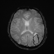

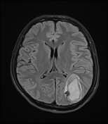
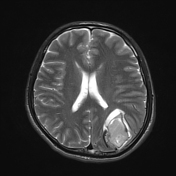
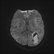
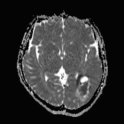
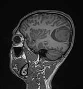
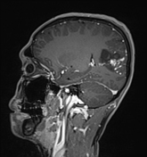
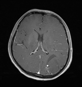
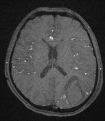
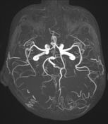
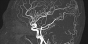
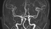

MRI of the brain shows a hemorrhage in the left parieto-occipital region measuring approximately 40 x 50 x 42 mm, with blood in the late subacute stage (high signal on T1W and T2W, low signal on GRE, and restricted diffusion), along with surrounding edema. Adjacent to the hematoma, there are tortuous dilated vascular structures with low signal on T2W, suggestive of a cerebral arteriovenous malformation (cAVM) with a nidus measuring 14 x 9 mm, enhancing after contrast administration. MRA and post-contrast T1FS reveal feeding arteries from branches of the middle cerebral artery and left posterior cerebral artery, with venous drainage into the cortical veins of the parietal region and into the superior sagittal sinus.
Case Discussion
This is a case of non-traumatic intracerebral hemorrhage in an atypical location not commonly associated with hypertension (i.e., not in the basal ganglia). Hemorrhage in this location raises suspicion of several possible causes, such as cerebral vascular malformations (ruptured AVM or cavernoma), intracranial venous thrombosis, or brain tumor.
In this patient, the MRI findings and clinical symptoms are consistent with a cerebral arteriovenous malformation (AVM) with hemorrhagic complications.


 Unable to process the form. Check for errors and try again.