
Presentation
Hard, painless left-sided breast lump
Patient Data
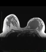

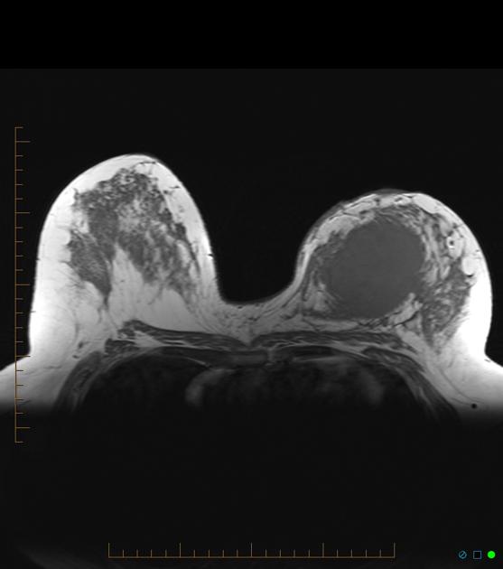
A well-defined ovoid mass lesion in the left breast. Mass is not particularly T2 hyperintense part from the central portion although there is considerable "edema" in adjacent breast parenchyma.
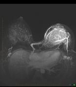
MIP of dynamic series showing a dramatic increase in blood flow to the entire left breast. Note recruitment of right internal mammary vessels.
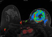
CAD color flow map showing red, malignant ("wash out") peripheral enhancement.
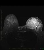
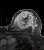
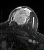
Peripheral enhancement with central non-enhancement corresponding to the T2 hyperintensity indicative of central necrosis. Note enlarged enhancing internal mammary node (arrowed). Enlarged, enhancing level 1 axillary lymphadenopathy (arrowed).
3D volume rendered reconstruction of abnormal enhancement of the left breast showing the size and extent of tumor involvement on this anterior view (note the subtracted view of the tumor alone in the left corner). Volume of tumor calculated as 268 cc.
Case Discussion
MRI is the investigation of choice for staging breast cancer ie determining size and number of tumors in the breasts (both ipsi and contralateral). Blood flow characteristics are also useful to determine response to neoadjuvant chemotherapy prior to surgical excisison.


 Unable to process the form. Check for errors and try again.