
Presentation
Repeated chest infection, cough and shortness of breath.
Patient Data
Age: 40 years
Gender: Male
From the case:
Bronchial carcinoid


Download
Info
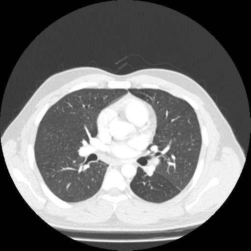
1.6 x 2.9 x 1.5cm well defined heterogeneously enhancing soft tissue lesion is seen in left lower lobe bronchus causing obstructive atelectasis, consolidation and bronchiectasis in the left lower lobe.
No collapse or consolidation is seen in the right lung.
Few subcentimetre prevascular and left paratracheal lymph nodes are seen.
Visualised sections of the upper abdomen a normal liver, pancreas, spleen, both kidneys and gall bladder.
No pathology is seen in bones.
Case Discussion
Bronchoscopy was done which showed mass in left lower lobe bronchus. Biopsy results confirmed carcinoid tumour of left lower lobe bronchus.


 Unable to process the form. Check for errors and try again.